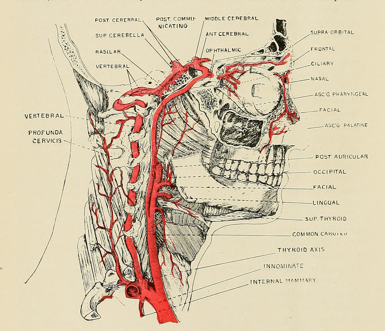
Arteries In Neck Labeled : The Neck Arteries 3 Pages 1918 Human Anatomy Illustration - Find this pin and more on anatomy and pysiology block 2by tonna brinson.
byAdmin•
0
Arteries In Neck Labeled : The Neck Arteries 3 Pages 1918 Human Anatomy Illustration - Find this pin and more on anatomy and pysiology block 2by tonna brinson.. The latter is less invasive, but some research is showing that this method may have a higher risk of complications. The neck is supplied by arteries other than the carotids. The right and left subclavian arteries give rise to the thyrocervical trunk. A pathological study to show the pattern of arterial involvement. The neck is arbitrarily subdivided into two triangles by the.
The neck is arbitrarily subdivided into two triangles by the. Labeled diagram of the arteries of the head and neck. Neck dissection removes potential or proven metastases to cervical lymph nodes. A blockage in one of the carotid arteries can be cleared either by endarterectomy or carotid angioplasty. The latter is less invasive, but some research is showing that this method may have a higher risk of complications.
34 Cat Veins And Arteries Diagram - Wiring Diagram List from lh5.googleusercontent.com Neck dissection removes potential or proven metastases to cervical lymph nodes. So hopefully that's given you a good overview of the arterial supply to the lower limb. A person with neck swelling has enlargement of the soft tissues that covers the neck. The largest arteries in the neck are the common carotids. Is a delicate, subcutaneous muscle separating the skin from the deeper anterior muscles of the neck. The growing plaque may eventually narrow the carotid artery, known as stenosis, and can lead to a stroke. The coronary artery and the circumflex artery are responsible for delivering oxygenated blood to the heart and break it is also the biggest artery in the human body. The hypoglossal nerve has been displaced downward in this preparation (lingual artery labeled at center left).
They ascend through the neck without branching before.
Labeled diagram of the arteries of the head and neck. Always read the nutrition labels in the foods you buy. The principal arteries are the carotid and subclavian arteries. Carotid artery disease is when fat accumulates and blocks the blood flow of your neck arteries (carotid arteries).your carotid arteries supply your brain with blood rich in oxygen. Find this pin and more on anatomy and pysiology block 2by tonna brinson. This diagram with labels depicts and explains the details of neck arteries. Vertebral arteries (arteria vertebralis) next to your spine which become basilar artery (arteria basillaris) and brings blood to the back of the brain. The principal arteries are the carotid and subclavian arteries. The left common carotid artery and left subclavian artery arising directly from the arch of the aorta to supply similar territories on the left side of the body. Master your knowledge about the anatomy of the nerves and arteries of head and neck at kenhub. The neck is supplied by arteries other than the carotids. The superior gluteal artery and the inferior gluteal artery supply the gluteal region, but we'll take a look at the branches of the internal iliac artery in more detail in separate tutorials. The growing plaque may eventually narrow the carotid artery, known as stenosis, and can lead to a stroke.
The left common carotid artery and left subclavian artery arising directly from the arch of the aorta to supply similar territories on the left side of the body. Arteries of the femoral n… category: Answer to label the arteries of the neck in the ct angiogram. Bodytomy provides a labeled iliac artery diagram to help you understand the anatomy and function of the common iliac. This entry was posted in anatomy, body parts, system and tagged anatomy of arteries, anatomy of artery, arteries, arteries anatomy, arteries chart, arteries diagram, arteries diagram with labels, arteries explained, artery.
nemfrog - Arteries of the neck. Applied Anatomy: Designed... from 78.media.tumblr.com These arise from the brachiocephalic trunk on the right side and directly from the arch of the aorta the internal carotid arteries are predominant contributors to the intracranial blood supply. Variable in extent, the platysma typically spans the space between the superior margins of pectoralis. Find this pin and more on anatomy and pysiology block 2by tonna brinson. From this trunk, several vessels arise, which go on to supply the neck. The first branch of the thyrocervical trunk is the inferior thyroid artery. Temporary blindness in one eye, usually caused by a fragment of. Is a delicate, subcutaneous muscle separating the skin from the deeper anterior muscles of the neck. The right and left subclavian arteries give rise to the thyrocervical trunk.
The left common carotid artery and left subclavian artery arising directly from the arch of the aorta to supply similar territories on the left side of the body.
A person with neck swelling has enlargement of the soft tissues that covers the neck. The superior gluteal artery and the inferior gluteal artery supply the gluteal region, but we'll take a look at the branches of the internal iliac artery in more detail in separate tutorials. Always read the nutrition labels in the foods you buy. Simple labelled illustration depicting the general pathways for the major arteries of the head and neck. The principal arteries are the carotid and subclavian arteries. The principal arteries are the carotid and subclavian arteries. It is bound laterally by the carotid arteries, superiorly by the hyoid bone, and inferiorly by. The growing plaque may eventually narrow the carotid artery, known as stenosis, and can lead to a stroke. Depiction of the neck with muscles and arteries shown. Carotid artery disease is when fat accumulates and blocks the blood flow of your neck arteries (carotid arteries).your carotid arteries supply your brain with blood rich in oxygen. Answer to label the arteries of the neck in the ct angiogram. Anterior triangle of the neck in detail. From this trunk, several vessels arise, which go on to supply the neck.
Ninja nerds, join us in this video where we use a human anatomy model to show the arteries of the join us in this video where we discuss the blood vessels of the head and neck and go into great dr mitusha verma explains the concept of 3d arterial spin labelling that helps in improving your. It carries blood from the left ventricle to the coronary arteries. It is located on every side of the neck and is a large triangular space, with its apex pointed downwards and base pointed upwards and in front of… it's partially concealed by the posterior edge of the sternocleidomastoid. The principal arteries are the carotid and subclavian arteries. Master your knowledge about the anatomy of the nerves and arteries of head and neck at kenhub.
Arteries Head Neck 2 Sided 1933 Human Anatomy Illustration ... from i.pinimg.com Labeled diagram of the arteries of the head and neck. The right and left subclavian arteries give rise to the thyrocervical trunk. The largest arteries in the neck are the common carotids. From this trunk, several vessels arise, which go on to supply the neck. A neck swelling can also occur as accumulation of fluid, lymph, or inflammatory, or tumor cells in an area unde… openstax cnx. The growing plaque may eventually narrow the carotid artery, known as stenosis, and can lead to a stroke. It supplies the thyroid gland. Ninja nerds, join us in this video where we use a human anatomy model to show the arteries of the join us in this video where we discuss the blood vessels of the head and neck and go into great dr mitusha verma explains the concept of 3d arterial spin labelling that helps in improving your.
A pathological study to show the pattern of arterial involvement.
This diagram with labels depicts and explains the details of neck arteries. Left axillary a left vertebral a left carotid sinus right subclavian. Is a delicate, subcutaneous muscle separating the skin from the deeper anterior muscles of the neck. The neck is supplied by arteries other than the carotids. Cholesterol plaque may slowly build up in the carotid artery wall, over decades. They ascend through the neck without branching before. Neck dissection removes potential or proven metastases to cervical lymph nodes. It carries blood from the left ventricle to the coronary arteries. The principal arteries are the carotid and subclavian arteries. It is located on every side of the neck and is a large triangular space, with its apex pointed downwards and base pointed upwards and in front of… it's partially concealed by the posterior edge of the sternocleidomastoid. The latter is less invasive, but some research is showing that this method may have a higher risk of complications. It supplies the thyroid gland. Variable in extent, the platysma typically spans the space between the superior margins of pectoralis.
This entry was posted in anatomy, body parts, system and tagged anatomy of arteries, anatomy of artery, arteries, arteries anatomy, arteries chart, arteries diagram, arteries diagram with labels, arteries explained, artery arteries in neck. The growing plaque may eventually narrow the carotid artery, known as stenosis, and can lead to a stroke.